thedarkrider on BudgetPixel
@thedarkrider · 4/16/2026
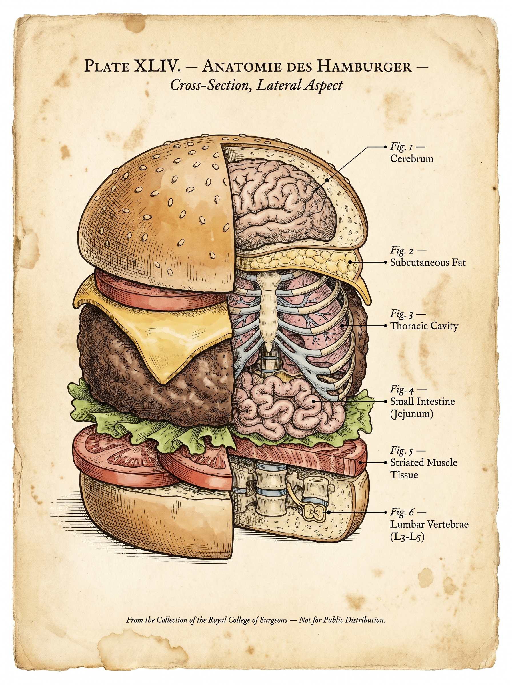
Plate XLIV: Anatomie des Hamburger — The Burger That Thinks It’s Alive
Tags: hamburger anatomy, cross-section, vintage medical illustration, educational plate, food anatomy, brain and organs, thoracic cavity, small intestine, labeled diagram, culinary art, anatomical illustration, retro science art, medieval medical engraving, educational anatomy image
AI Model: Nano Banana Pro
Prompt: A detailed anatomical cross-section illustration in the precise style of a 19th century medical textbook plate — fine crosshatched ink linework on aged yellowed parchment paper with subtle foxing stains at the edges, the meticulous hand-drawn quality of Gray's Anatomy or a Vesalius engraving. The subject is a realistic hamburger rendered in three-quarter view with the right half cut away cleanly to expose a vertical cross-section, as if sliced with a surgical blade. The exterior left half looks like a completely normal appealing cheeseburger — golden sesame seed bun, crisp green lettuce, red tomato, melted cheese, thick beef patty, all rendered with appetizing realism in muted hand-tinted color washes over the ink drawing. The exposed right half reveals human anatomical structures perfectly nested inside each layer in place of the food, rendered in the exacting detail of a medical illustration with precise stippling and crosshatching. The top bun's interior contains a complete human brain — cerebral folds and fissures drawn in meticulous detail, the gyri and sulci fitting perfectly within the dome of the bun, a thin layer of sesame seeds still visible on the outer surface transitioning seamlessly into grey matter beneath. A fine ink leader line extends to a serif label reading "Fig. 1 — Cerebrum." Below the brain, the cheese layer is a thin membrane of adipose tissue labeled "Fig. 2 — Subcutaneous Fat." The thick patty layer contains a detailed rib cage cross-section — curved ribs encasing two pink-grey lungs with visible bronchial branching, the tissue rendered with the wet organic texture of an actual anatomical specimen, labeled "Fig. 3 — Thoracic Cavity." The lettuce layer peels back to reveal tightly coiled loops of small intestine packed with the dense efficiency of actual bowel, the mesentery faintly visible connecting the loops, labeled "Fig. 4 — Small Intestine (Jejunum)." The tomato slices show exposed striated skeletal muscle fibers in cross-section — the fascicle bundles clearly delineated with the characteristic banding pattern visible at the cut surface, labeled "Fig. 5 — Striated Muscle Tissue." The bottom bun contains three lumbar vertebrae stacked with intervertebral discs between them, the spinal canal visible in the center with a tiny cross-section of the spinal cord inside, labeled "Fig. 6 — Lumbar Vertebrae (L3-L5)." All labels are printed in an elegant serif typeface in the style of 19th century anatomical plate annotations, connected to their structures by fine straight ink leader lines with small terminal dots. At the top of the page, centered, reads "PLATE XLIV. — ANATOMIE DES HAMBURGER — Cross-Section, Lateral Aspect" in small elegant serif capitals. At the bottom, small text reads "From the Collection of the Royal College of Surgeons — Not for Public Distribution." The color palette is the muted hand-tinted wash of a Victorian medical illustration — soft pinks and reds for muscle and organ tissue, grey-brown for the bun exterior, pale yellow for fat, blue-grey for bone, all over the warm parchment base. No border, no frame, edge to edge.